Subcostal (Subxiphoid) Four Chamber View
Ok, so let's start off with one of the easiest views, the Subcostal Four Chamber view. You are likely familiar with this view already as it is part of the FAST exam. It provides a great image of the entire heart (but remember to zoom in to at least 20cm or you won't find it!). This view is ideal for looking for pericardial effusions, global function of the heart and is especially great in COPD patients whose large lung fields may obscure other cardiac views. Very heavy patients or ones with distended abdomens may provide suboptimal images (or no image at all!).
Probe Placement
Probe placement for the Subcostal view is easy, once you know the correct position. Place the transducer with the marker to the patient's RIGHT side, and lay it flat just beneath the xiphoid. Apply reasonable pressure to the subxiphoid area, and if possible, ask the patient to take a deep breath. This will move the heart inferiorly and give you a better view.
Subcostal probe placement (shoot subxiphoid and through the liver edge). Usually we point the marker to the patient's RIGHT.
 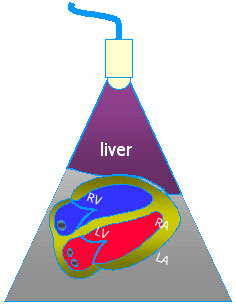
And here's what you'll see on the screen (note: the probe marker is to the patients LEFT shoulder in this shot (instead of to the right), which flips the heart on the ultrasound monitor)...It's ok to point the marker to either side you like, AS LONG AS you're consistent, and know WHY the heart appears as it does on your monitor with respect to your probe marker!)
As in the picture, the right side of the heart is at the top of the screen (as it is in just about every cardiac view...). You can decipher the ventricles from the atria I'm sure!
Onto our next view...the parasternal long axis
|