"Flank Pain"
Answer: Hydronephrosis
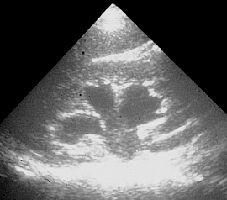
Bedside ultrasound demonstrated a normal left kidney and mild hydronephrosis of the right kidney. No stones were visualized in either renal pelvis.
Ultrasound of the patient’s fetus was also performed. The fetal heart rate was 147 and regular. The fetus’ head diameter was appropriate for the stated gestational age.
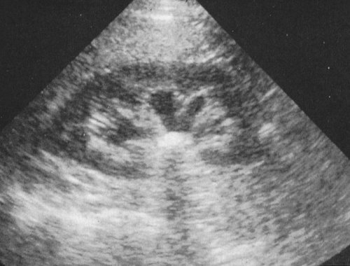
Hydronephrosis Review
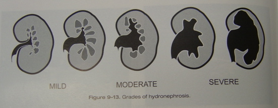
Hydronephrosis has been incidentally found at autopsy in all age groups 3% of the time. It is defined as a dilation of the renal pelvis and calyces.
This abnormal finding is usually a result of distal obstructive disease. Potential causes of obstructive disease include any anatomic, functional, or metabolic derangement halting flow
from the renal pelvis to the distal urethra. In children, the most common causes are reflux and ureteropelvic junction obstruction. Calculi are the most common cause in young adults.
Older men may develop hydronephrosis as a result of obstruction from prostate cancer or BPH. Finally, women encounter problems in conjunction with pregnancy and gynecologic cancers.
Graphic from Ma/Mateer, Emergency Ultrasound
Researchers have shown that with bedside ultrasound and the correct clinical setting, patients such as this one may be managed as an outpatient with close follow-up. Bedside renal
ultrasound performed by ED physicians has proven to be as sensitive as CT (86% vs. 83%, respectively) and nearly as specific (83% vs. 92%, respectively) for the detection of hydronephrosis.
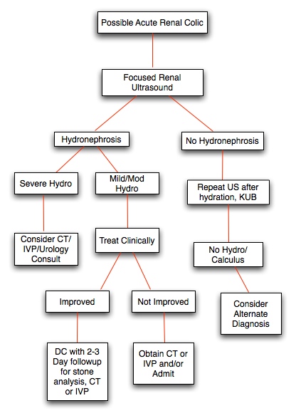
Per an algorithim from Ma/Mateer's text, patients with mild to moderate hydronephrosis may be discharged with followup if clinically improved. However, a patient with severe hydronephrosis
(as documented by ultrasound) should be managed with CT and urologic consultation.
Management flowsheet dervived from Ma/Mateer, Emergency Ultrasound
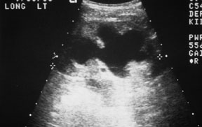
Modereate/Severe Hydronephrosis
Interestingly, pregnancy can produce a degree of physiologic hydronephrosis. This physiologic hydronephrosis will appear similar in ultrasound images to a mild obstructive uropathy. Several theories have been proposed with respect to the development of physiologic hydronephrosis. These include: increased vascular volume, decreased prostaglandin activity leading to decreased ureteral peristalsis, hormonal changes leading to renal calyce relaxation and dilation, and pelvic floor muscle relaxation leading toincreased incidence of ureteral mechanical obstruction at the pelvic brim.
Dilated Renal Calyces
REFERENCES:
1. http://www.emedicine.com/med/topic1055.htm
2. Kartal, M. Prospective validation of a current algorithm including bedside US performed by emergency physicians for patients with acute flank pain suspected for renal colic. Emer Med J. 2006 May; 23(5): 341-4.
3. Gaspari, RJ. Emergency ultrasound and urinalysis in the evaluation of flank pain. Acad Emerg Med. 2005 Dec: 12(12): 1180-4.
4. Brenner, Barry. Brenner & Rector’s The Kidney: Saunders, 2003.
5. Ma OJ,Mateer J. Emergency Ultrasound: McGraw Hill, 2003.
Back Home
|