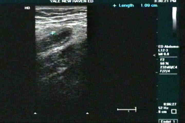
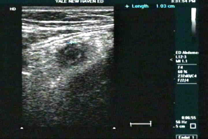
Ultrasound Case of the Week #15
If you guessed an appendicitis, you're right. The CT revealed a perforated appendix with abscess formation. The patient received IV antibiotics for several days and removal of the abscess is planned in several weeks.
Two other images of appendicitis from New Haven ED


Appendicitis and Ultrasound
Ultrasound for the diagnosis of appendicitis was first described by Julien Puylaert in 1986. His technique, "graded compression sonography" was discussed in a paper including 60 subjects suspected of having an acute appendix. It was found, in conjunction with other studies, that the use of ultrasound in the diagnosis of appendicitis can reduce negative laparotomy rates to 10%, a figure much improved over clinical exam alone (this doesn not include CT of course). The diagnosis is entirely dependent on visualization of the appendix that is inflamed : "a blind-ended noncompressible, aperistaltic tube, arising from the tip of the cecum with a gut signature." (Rumack)
The acutely inflamed apppendix may be recognized by several key features :
1. Greater than 6-7mm diameter of the appendix
2. Round shape of the appendix (as opposed to ovoid)
3. Non-compressibility of the appendix (a normal appendix is easily compressible and <3mm in diameter)
4. Visualization of an appendicolith is diagnositic of appendicitis.
5. Color doppler may reveal hyperemia about the appendiceal wall (due to inflammation).
Additional considerations:
It has been described that that appendix is more difficult to visualize in women as it may slide down into the true pelvis, due to larger pelvic inlet size. Performance of transvaginal ultrasonography has revealed an inflammed appendix in several cases.
Thin patients are much better suited for ultrasounding the appendix than are obese pateints (for obvious reasons).
The sensitivity of the identification of appendicitis decreases with perforation, though one may see:
1. loculated pericecal fluid
2. phlegmon or abscess
3. prominent pericecal or pperiapppendiceal fat
4. circumfrential loss of the submucosal layer of the appendix
In summary, if you have a thin patient who you are suspicious may have appendicits, give it a try and look for the appendix that is dilated, circular and noncompressible! Then you can confirm it with the CT you ordered, as I know as well as you that no one at Richland is going to the OR without one!
Thanks for looking!
Back Home
|
|
|